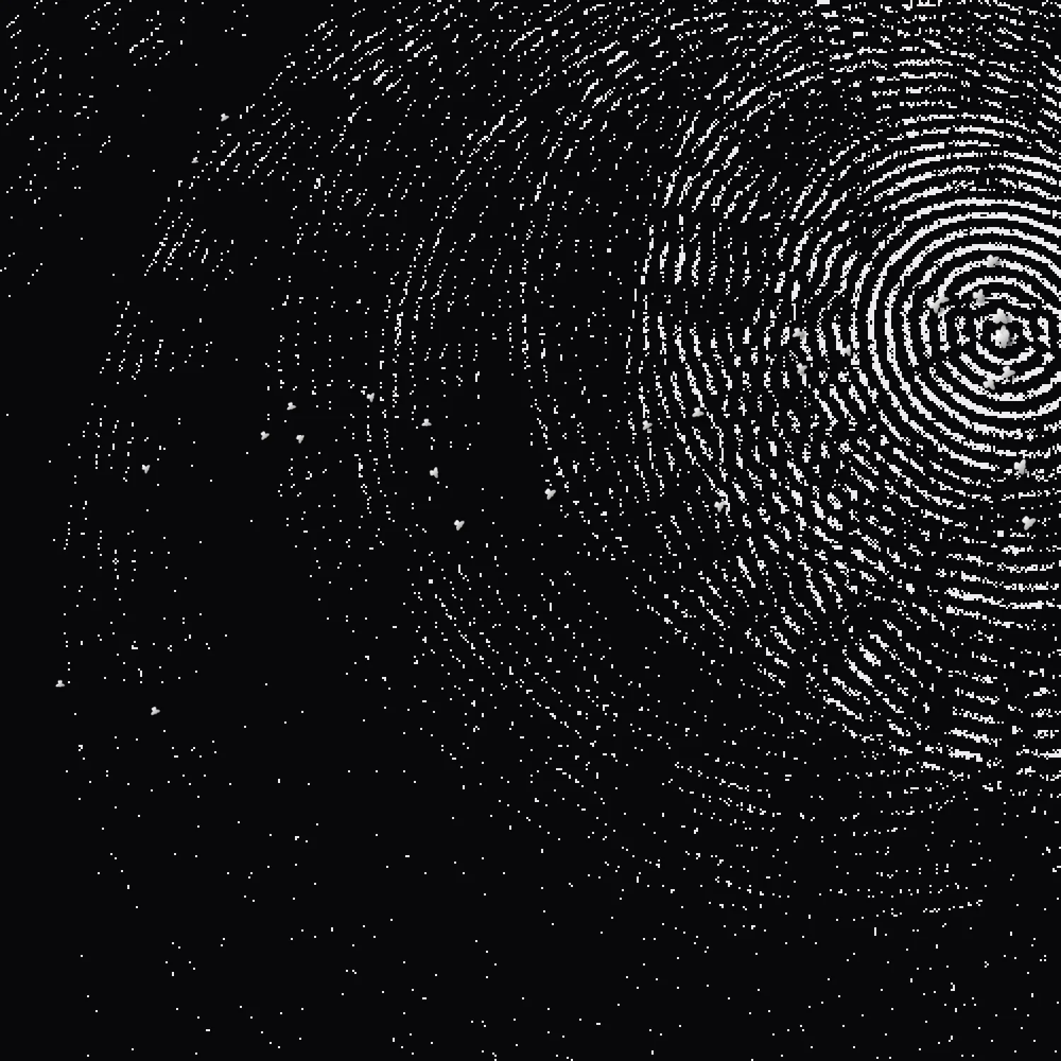
When you enter a very dimly lit room, at first the details are muted, and then slowly the objects and space around you start to reveal themselves. You see the edge of a table, the glint of your toaster, folds in a curtain, the outline of your own hand. Deep in the rod cells at the back of your eye, a protein called rhodopsin is catching that faint light and turning it into a signal you can see.
Earlier this month the 2026 Nobel Prize in Physiology or Medicine went to Karl Deisseroth, Peter Hegemann and Georg Nagel "for their discoveries concerning light-gated ion channels and optogenetics", a technique that lets you turn specific neurons on and off with pulses of light. This Nobel winning technique was made possible due to a type of rhodopsin protein that came not from the human eye, or from any animal at all, but instead from a single-celled green alga. The most precise tool neuroscience has for controlling the neurons in your brain came from a creature that doesn’t even have one.
The discovery process of how this field-opening protein made its way from the soil to the brain doesn’t follow a straight and predictable journey. Instead, it unfolds as a series of seemingly disparate scientific contributions and curiosities. The path runs from a microbe that lives on sunlight in brine, through frog eggs in Frankfurt, an open catalogue of genes in Japan, over thirty years and through fields that had nothing to do with neuroscience at all. It’s decidedly non-linear.
This is true of many important scientific discoveries. A lot of the critical pieces on the discovery trail could look modest or unrelated when they were initially developed. They lay frozen in the collective scientific consciousness, sometimes for decades, until someone with a different perspective picked them up and asked questions far from those that led to their inception.
This piece traces the discovery lineage of optogenetics, and shows how great scientific work grows the way living things do. Flowing across time, people, instruments, branching and borrowing, advancing through detours, delays, serendipity, and exchange. Along the way, the significance of a finding appears less in the question it answered than in how many directions it opened beyond itself. I call this the fertility of an idea.
From pink salt ponds to frog eggs
Our journey begins with the salt-loving microbe, Halobacterium, which thrives in brine almost too salty for anything else to live in. These microbes grow so densely that they transform bodies of water into dreamy pink and purple pools. It was in 1971 when Dieter Oesterhelt and Walther Stoeckenius, studying this microbe, discovered bacteriorhodopsin.

1971-1982Turning sunlight into energy
Bacteriorhodopsin sits in patches of the microbe's membrane, coloured purple by the retinal – a light-absorbing molecule made from vitamin A – that it holds. Within a couple of years, Oesterhelt and Stoeckenius had shown that it uses light to pump protons across the cell membrane. Each time the light hit, it pushed a proton out of the cell, which was a simple way of turning sunlight into usable energy.
Soon enough, 6 years later, Yasuo Mukohata's group in Osaka found signs of a second light-driven pump in the same microbe, Halobacterium. The protein, later named halorhodopsin, was first assumed to be a sodium pump, but in 1982 Brigitte Schobert and Janos Lanyi showed it actually moves chloride. At the time, both bacteriorhodopsin and halorhodopsin belonged to the category of research dealing with how microbes make energy. They weren’t yet seen as potential tools for neuroscience, which would ultimately prove to take some time.
1975-1990Resolving structure from noise
Even before the cross over into neuroscience, bacteriorhodopsin had already literally changed how biologists see proteins. It started with a problem: the electron microscope tends to destroy the very samples that it could give scientists the power to see. But in the purple membrane of the bacteriorhodopsin’s microbe, this protein happens to pack itself into a naturally regular lattice, repeating the same unit thousands of times. In 1975 (4 years after the discovery of bacteriorhodopsin) Richard Henderson and Nigel Unwin realized that they could use this lattice to their advantage. They turned the microscope’s beam down so low that each individual image was mostly noise, letting the regularity of the lattice do its work, and then combined the signal from thousands of identical copies into one clear picture.
Their model was the first three-dimensional picture of a protein made with an electron microscope, and it showed seven helices packed side by side, each one crossing the membrane. Henderson kept refining the approach, and by 1990 had a near atomic-resolution model.
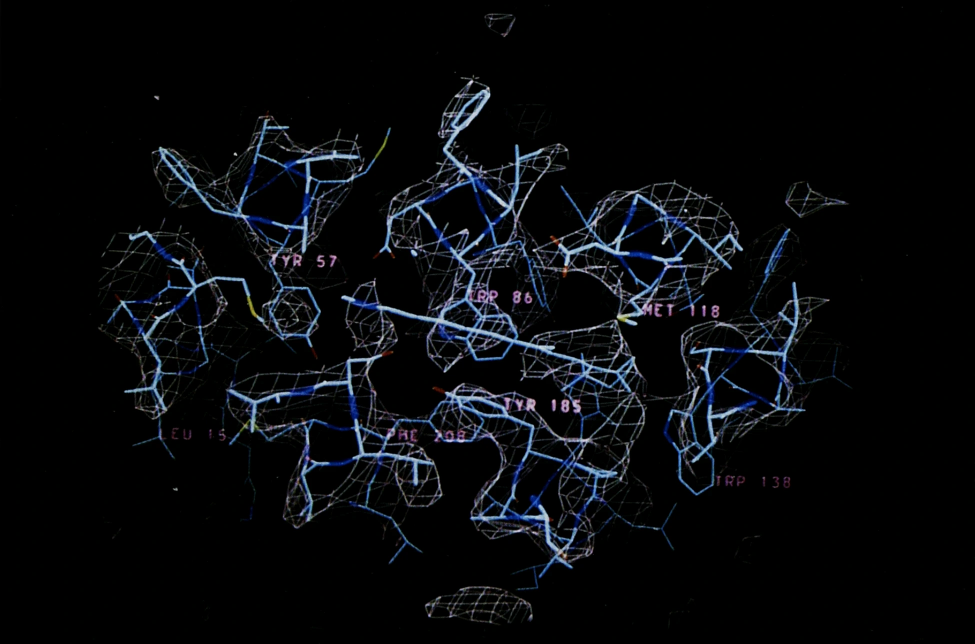
This very same logic of averaging many noisy images of identical molecules grew into cryo-electron microscopy, which won the 2017 Nobel Prize in Chemistry. The discovery of bacteriorhodopsin in 1971 set off a chain no one could have foreseen.
And while bacteriorhodopsin was opening up a new way of seeing in biology, another line of inquiry was forming around what more these light-driven rhodopsin proteins could do and where else they might be found.
1985-2001Could the sensor be the switch?
Peter Hegemann, who had worked on the light-driven protein halorhodopsin as a PhD student in Oesterhelt's lab, moved in 1985 to work with Kenneth Foster, who had just shown that the green alga Chlamydomonas senses and navigates by light using (a not yet specified) rhodopsin. In 1991, using a suction-pipette technique first developed in Moscow on a different alga, Hegemann's group recorded currents in the alga that appeared almost the instant the light did.
This speed was of particular interest because the rhodopsin in your eye isn’t nearly as fast as this. When light enters your eye, the rhodopsin works as a sensor rather than a switch and when it catches light it passes the news to a relay of other proteins, each one multiplying the message until there’s enough to change the cell’s electrical state. Every handoff along this relay takes time.
The currents in the alga came too quickly for anything like that…so how did they work? The simplest explanation was that there was no relay at all, and that in the alga the light sensor and the switch were the same protein. Instead of a sensor relaying information along, the rhodopsin would itself act as a light-gated channel that once triggered, would open up, letting charged particles flow through. This was a pretty compelling idea, however, no one had yet found a protein that displayed these properties.
1995-2003A new channel of discovery
Unbeknownst to them yet, the protein they were looking for was sitting in an open algal gene sequence library, published by Japan's Kazusa DNA Research Institute. Roughly thirty years after bacteriorhodopsin's discovery, Suneel Kateriya in Hegemann's group searched that catalogue for genes resembling bacteriorhodopsin. He found two candidates in the alga Chlamydomonas. Groups in Texas and Japan found the same two genes at almost the same time, but what nobody had yet shown was whether these proteins worked like light-gated channels that so far existed only as an idea.
As it happened, the way to find out and test the two candidates had already been developed years earlier to answer another question. It was in 1995 when Georg Nagel and Ernst Bamberg wanted to know how hard bacteriorhodopsin could push protons against a voltage – measuring the strength of its light-driven pump. Their solution was to express bacteriorhodopsin in frog oocytes, the immature egg cells of a frog, using a trick borrowed from developmental biology that had already crossed over into neuroscience. In solving their own problem, they also showed that a microbial rhodopsin could work in something as complex as an animal cell. Six years later, in another twist along the lineage of discovery, their work could be applied to a question about algal rhodopsins.
Hegemann, already familiar with this work in frog oocytes, and having worked with Bamberg on halorhodopsin in the 1980s, teamed up with both Nagel and Bamberg and sent them the two candidate genes that Kateriya had identified. Could these be the proteins they were looking for?
When Nagel and Bamberg expressed these two genes in frog oocytes, the proteins indeed turned out to be channels that opened in response to light! They named the first channelrhodopsin-1 and published it in 2002, and a year later described the second, channelrhodopsin-2, which let positively charged ions flood into the cell whenever light hit it. In that 2003 paper they also tried it in mammalian cells, and suggested it could be useful for manipulating the membrane potential of mammalian cells.
These were biophysicists interested in a very particular research area: how proteins move ions across membranes. Despite such a seemingly narrow focus, they had, perhaps without quite meaning to, opened the door to a field that didn’t yet exist.
From frog eggs to firing neurons
Over in neuroscience, researchers really wanted the ability to switch one type of neuron on or off without triggering any other nearby cells. By the late 1990s some researchers were wondering whether light might be the answer.
One of the early genetic light-control attempts came in 2002 when Gero Miesenböck's group made neurons light-sensitive by giving them a set of genes borrowed from the light-sensing cells of fruit flies. They were the first to prove that the idea was possible. But the system they built was, in effect, similar to the relay from your eye: they had several proteins working together, each passing the message on to the next, and every handoff taking time. What neuroscience was still lacking at this time was something simple, fast, and self-contained.
2004-2006Firing at the flash of a light
Over at Stanford, Ed Boyden had read the channelrhodopsin paper and together with Karl Deisseroth who was just setting up his own lab, asked Nagel for the gene.
Feng Zhang, a graduate student working with Deisseroth, used a virus to deliver this gene into rat neurons growing in a dish. Then came the pivotal experiment where Boyden pulsed blue light onto one of the prepared neurons and recorded a fast, precise electrical spike in response to most of the flashes. These findings were published in 2005 and within a few years, channelrhodopsin became the basis of a new way to control neural activity.
As it turned out, they weren't the only ones putting channelrhodopsin into neurons. When the foundations and preconditions of an idea are in place, the same discovery has a habit of appearing in more than one place at once. The sociologist Robert Merton called these “multiples” and thought they were the rule rather than the exception.
So it’s not so surprising then, that in Detroit, Zhuo-Hua Pan at Wayne State University had been trying to restore sight by making the surviving cells of a degenerated retina respond to light. In early 2004, months before Boyden's experiment, he observed retinal neurons – carrying the very same channelrhodopsin – respond to light. He struggled to get his work published, before his paper was finally accepted in 2006.
While Pan waited for his paper to be accepted, the technique was already spreading. Within four months of the 2005 Stanford paper, labs in Ohio, Frankfurt and Sendai had channelrhodopsin working in chick spinal cords, worms and mouse brain slices. In 2006, Deisseroth and his colleagues gave the growing approach a name, optogenetics: control by light, written into cells by their genes.
2007-2010Blue to fire, yellow to fall silent
The salt pond had one more gift to give us. Halorhodopsin, the second protein found in 1977 in Osaka, turned out to be very valuable 30 years later, when a version from a related microbe became the off switch. This turned out to be another “multiple” found independently by Ed Boyden’s lab and Deisseroth’s, letting neuroscientists silence neurons with yellow light.
Soon, light could be delivered through thin optical fibres into the brains of mice that were free to move, and in 2010, the journal Nature Methods named optogenetics its Method of the Year.
The scientific cascade of contributions that paved the way to optogenetics spent many years in a seemingly distant space, beginning with microbiologists studying light-driven pumps in a purple-membraned microbe. This thread of scientific intuition progressed through researchers studying a green alga who found that it responded to light faster than an animal eye can, into an open catalogue of algal genes that let them find the protein responsible. Soon after, biophysicists showed that these algal genes made light-gated channels, and from there, neuroscientists put the channels into neurons, and so, a new field was born.
We had finally arrived at a field that today is the subject of much acclaim and investment, following a path that wound and wandered.
Discovery is relational
Back in 1975, Henderson and Unwin could only see bacteriorhodopsin by stacking thousands of noisy images until a distinct shape began to come through. With more images and better instruments, the image grew sharper. Today, the methods that grew from their work give us atomic resolution, still reliant on the same principle that many imperfect images, together can yield a signal that no single one of them could provide.
The history of optogenetics follows a similar logic. Taken one at a time, each scientific contribution along its lineage of discovery can seem isolated and obscure, their potential not yet clear. But when you assemble them all together, a complex structure resolves.
Discovery is relational. It comes into focus the way a protein does, its shape emerging only as each lattice of inquiry is layered over the last. Below is the lineage of optogenetics, resolved into a single image.
It shows ideas moving through microbial biochemistry, structural biology, algal photobiology, gene sequencing, neuroscience, and eventually the clinic. These ideas are carried by many independent hands across the globe, most of them working on separate questions, not yet aware of how much their paths crossing would come to matter. It’s dramatically nonlinear, unpredictable, and organic.
The history of major scientific discovery is often compressed into Nobel winning stories about a few geniuses and some perfectly timed breakthroughs, but the actual lineage is more distributed, less intentional and constantly evolving. Optogenetics wasn't discovered so much as it emerged and continues to grow in a fertile research ecology, through the people and materials crossing between fields.
It’s also never ending. Most of the right-hand side of the figure continues past the edge of where the field of optogenetics initially opened up, and goes on to trace its continued growth. With these new tools in hand, researchers have a huge playground of possibility before them. They conducted experiments like activating one type of striatal neuron showing that they could relieve parkinsonian movement issues in mice, or using light to stop seizures in epileptic mice, and reactivating neurons that stored lost memories in mice modelling early Alzheimer’s disease.
Meanwhile, new channelrhodopsins keep popping up from sequencing data, and more recently machine learning, directed towards new lines of inquiry. In 2021, a man with retinitis pigmentosa partly regained his sight, and just this month, the full results of that trial reported that 7 out of 10 patients became more sensitive to light.
By tracing what led to the discovery of optogenetics, it’s clear that there was no single, privileged moment of insight at the start. The critical judgments were scattered across the whole pipeline, made by different people for different reasons. A successful scientific contribution in that sense isn’t only one that produces an ideal result with obvious value, but one that leaves the world with more possible futures than it had before. This is what the fertility of an idea looks like in practice.
An interesting question starts to emerge: how do we evaluate the threads of a successful discovery lineage? How do we identify and measure what makes a research ecology fertile? Through this framing of fertility, relying on methods like counting paper citations starts to look increasingly outdated.
This distinction is especially critical now as we build models and machines to accelerate discovery. It’s tempting to point them at what we can already measure, the likeliest target, the cleanest data, the quickest route to a known goal. A system built that way might have had little use for a microbe in a pink salt pond or producing an open catalogue of algal genes with no distinct purpose attached, and yet those were some of the pieces that, decades later, invented a field. The optogenetics lineage figure above could only be drawn from the far end, once the lines had already met. That’s where I think the conversation about AI and scientific discovery needs to start, and where my next piece will begin.